Anti-neutrophil cytoplasmic antibodies (ANCA) were originally described in the context of chronic inflammatory diseases. Such antibodies were detected by indirect immunofluorescence (IIF) on ethanol-fixed neutrophilic granulocytes revealing a pattern of perinuclear staining (P-ANCA). Many years later it appeared that these antibodies were commonly present in patients with small-vessel vasculitis, although in these patients two distinct patterns were observed, i.e., the P-ANCA pattern and granular cytoplasmic staining (C-ANCA).
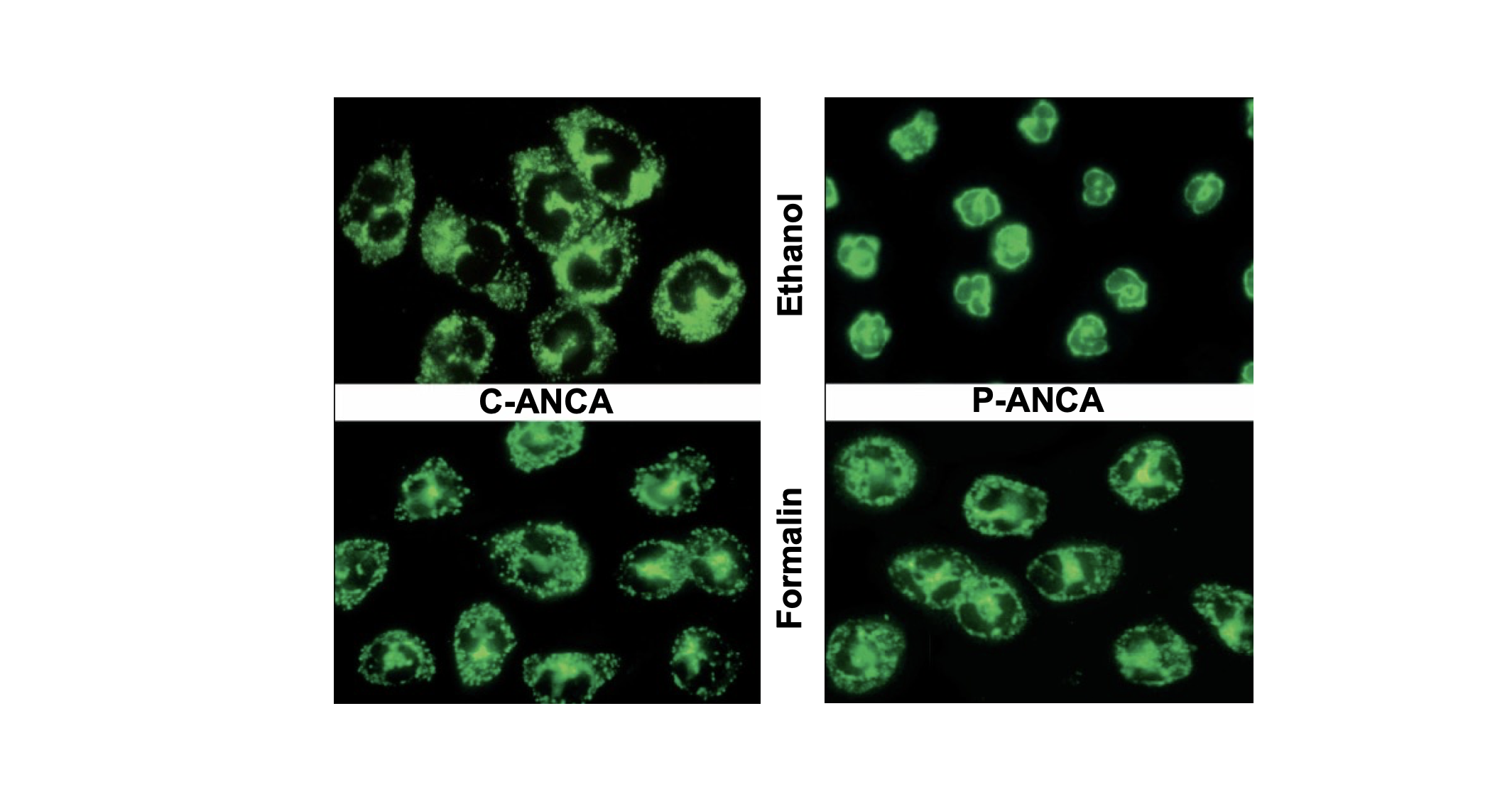
In contrast to the C-ANCA pattern, the P-ANCA pattern appeared to be an artefact due to the fixation with ethanol. Indeed, if the neutrophils were fixed with formalin, the sera that were identified with a P-ANCA pattern on ethanol-fixed neutrophils revealed a C-ANCA pattern on formalin-fixed neutrophils (Figure1).

Figure 1: ANCA-patterns of indirect immunofluorescence. PR3-ANCA reveal a granular fluorescence in the cytoplasm of ethanol-fixed (C-ANCA, upper-left) and formalin-fixed neutrophils (lower-left). MPO-ANCA, however, reveal a linear fluorescence around the de nucleus of ethanol-fixed neutrophils (P-ANCA, upper-right). This is a fixation artefact because formalin-fixed neutrophils reveal a granular fluorescence in the cytoplasm (C-ANCA, lower-right).
At the end of the ‘80s, the antigens recognised by ANCA in patients with small-vessel vasculitis were identified as myeloperoxidase (MPO) and proteinase 3 (PR3). This enabled the development of antigen-specific immuno-assays that were more specific for vasculitis than IIF. Due to the high sensitivity of IIF, the 1999 international consensus on ANCA testing prescribed that screening for ANCA should be performed by IIF with the follow-up of positive results by solid-phase assays of both MPO- as well as PR3-ANCA. In particular, the combination of C-ANCA/PR3-ANCA and P-ANCA/MPO-ANCA was highly suggestive of the so-called ANCA-associated vasculitis (AAV), consisting of granulomatosis of polyangiitis (GPA, previously Wegener’s granulomatosis), microscopic polyangiitis (MPA), and eosinophilic granulomatosis of polyangiitis (EGPA, previously Churg-Strauss syndrome).
Related: Antibody indices – a growing analysis in the diagnosis of CNS diseases
In the meantime, solid-phase assays gradually improved in terms of test characteristics, and multiple distinct assays, next to the original ELISA, entered the market. Since many of these systems were executed on fully automated analysers, laboratories increasingly abandoned IIF as a screening method, although solid evidence for this approach was not yet available. The multicentre study published in 2017, however, clearly revealed that the current immuno-assays for MPO- and PR3-ANCA outperformed the most optimal IIF strategy, i.e., the combination of ethanol- and formalin-fixed neutrophil substrates. This resulted in a revised international consensus stating that for GPA and MPA detection of ANCA by antigen-specific immuno-assays is to be preferred above screening with IIF. This consensus was subsequently extended to EGPA, although this was an eminent-based consensus because underlying data were lacking. The novel consensus also advised performing a confirmation test if results for MPO- or PR3-ANCA were only low-positive. Again, because antigen-specific assays outperform IIF, confirmation preferentially is performed with antigen-specific immuno-assays.
Based on the above, it might be conceived that ANCA IIF can be completely abandoned. However, several clinical settings may still profit from IIF. First, AAV or clinical mimics may be induced by drugs, one of which is cocaine. This may result in the generation of ANCA directed to human elastase (HLE), which reveals a P-ANCA pattern in IIF. Obviously, such sera may react negatively in antigen-specific assays for MPO- and PR3-ANCA. Many of these sera, however, are also positive for both MPO- as well as PR3-ANCA.
Secondly, ANCA as detected by IIF is not restricted to AAV, but is also encountered in inflammatory bowel disease, in particular ulcerative colitis, and autoimmune liver diseases like autoimmune hepatitis and primary sclerosing cholangitis. The antigens recognised by these ANCA are only poorly defined and, although some assays reveal an increased prevalence of PR3-ANCA in ulcerative colitis and primary sclerosing cholangitis, IIF seems to be the best method of detection.
Finally, ANCA IIF remains relevant in the 2022 classification criteria of the three distinct subtypes of AAV. These criteria are based on a dataset generated from 2011 – 2017, which is before the publication of the revised consensus on ANCA detection. In these criteria, P-ANCA is equivalent to MPO-ANCA while C-ANCA is equivalent to PR3-ANCA. It should be stressed, however, that these criteria should be applied in patients for which a diagnosis of small-vessel vasculitis has been established, but a classification for GPA, MPA, or EGPA has to be made.
Related: Guide to building pharma strategies in oncology
Since ANCA are not only used for diagnosis but also for follow-up of AAV patients, adequate positioning of IIF versus solid-phase assays also holds for this purpose. ANCA detection for follow-up may have three distinct intentions — first, to see if autoantibody levels decline after the start of immunosuppression; second, to determine the risk of relapse at the time clinical remission is achieved; and third, to predict a clinical relapse based on a rise in ANCA.
For the first objective, a decline can be observed equally well by both techniques as long as the end-point titer/level has been determined. For the second objective, it is important to know that this risk evaluation has originally been established by IIF, while more recently it was shown to be less predictive for solid-phase immunoassays. Finally, the added value of serial ANCA measurement for predicting a clinical relapse remains a matter of discussion. However, in general, solid-phase assays perform more accurately than IIF. For this objective, it is crucial that one-and-the same assay is used for follow-up because the results of distinct assays cannot be aligned.
Altogether, it is evident that ANCA IIF has played a very important role in the laboratory diagnostics of small-vessel vasculitis. Although ANCA IIF has recently been included in the criteria to classify the distinct disease entities of AAV, evidence for a multi-centre study clearly indicates that antigen-specific immuno-assays are better for the diagnostic work-up of these diseases. Positioning of IIF in other chronic inflammatory diseases cannot be replaced because the respective antigens are not well known.

Dr. Jan Damoiseaux is the Assistant Professor, Diagnostic Centre, NUTRIM School of Nutrition and Translational Research in Metabolism; Faculty of Health, Medicine and Life Sciences, Maastricht University Medical Center, Maastricht, The Netherlands. He will be speaking on “ANCA IF Pattern in Current Clinical Practice” at the Immunology Conference on August 18 at Medlab Asia 2023.
Learn more about Medlab Asia and Asia Health and click here to register for the event.
This article appears in Omnia Health magazine. Read the full issue online today.


