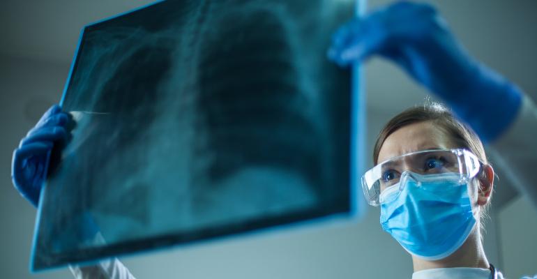
The typical findings of COVID-19 on chest radiography and computed tomography (CT) include bilateral, multifocal parenchymal opacities (ground-glass opacities with or without consolidation, and “crazy paving”). In most cases, the opacities are predominantly in the peripheral and lower lung zones, and several have rounded morphology.
However, these imaging findings are not pathognomonic for COVID-19 pneumonia and can be seen in other viral and bacterial infections, as well as with noninfectious causes such as drug toxicity and connective tissue disease. Most radiology professional organizations and societies recommend against routine screening CT to diagnose or exclude COVID-19.
The lungs are the most common site of infection in COVID-19, and progression to respiratory failure is the most common cause of death. In this brief summary we describe the role of thoracic imaging in COVID-19.
Chest radiography in COVID-19
Chest radiography is considered an appropriate initial imaging diagnostic test for most patients with lower respiratory tract infection, including those suspected of having COVID-19. The radiographic abnormalities in COVID-19 mirror those on computed tomography (CT), demonstrating bilateral, peripheral, and mid-lower-lung-zone-predominant consolidation.
However, in patients who have a high pretest probability of COVID-19, atypical findings such as diffuse interstitial changes or unilateral focal consolidation should not dissuade the radiologist from suspecting an infection, including COVID-19, as a possible diagnosis.
In patients with progressive disease, the density and extent of parenchymal changes typically increase over time. The severity of chest radiographic findings peaks 10 to 12 days after the onset of symptoms.
Unfortunately, most bacterial pneumonias also present as consolidation, and it is difficult to distinguish them from viral infections on chest radiography. The subtleties of rounded morphology and “crazy paving” associated with COVID-19 can only be appreciated on CT and not on plain chest radiographs. Cavitation within an airspace consolidation likely suggests a superadded infection.
Moreover, chest radiography has a high false-negative rate, especially in the early stage of infection, and should not be used as a screening tool to rule out COVID-19. In fact, baseline radiography has a lower sensitivity (69%) than initial reverse transcriptase polymerase chain reaction (RT-PCR) testing (91%).
Radiography 'through class' to avoid spreading the virus
During the pandemic, our hospital (as well as many others in the United States) has employed a method of obtaining portable radiographs in cases of confirmed or suspected COVID-19 through the glass wall of the patient’s room in the intensive care unit and in the emergency department.
With some minor technical modifications, the chest radiographs taken “through glass” are comparable to those obtained by the standard method. This technique has the potential to reduce the consumption of personal protective equipment by radiology technicians and to reduce the risk of machine contamination.

